To study the exclusive effect of khat chewing and rehabilitation therapy from khat addiction, ECG data was collected from healthy chewer subjects, control subjects and khat addicted subjects admitted to a rehabilitation centre. The variations in ECG signal features were extracted using signal processing techniques to analyse the changes in the cardiac activity. Figure 1 shows the general procedure used in this study.

General procedures of the study.
Study design
The effect of khat on heart activity
Sample selection and eligibility criteria
For the effect of khat on heart activity study, a quasi-interventional design approach was used. The subjects were selected based on a pilot survey conducted to identify appropriate subjects. The selection criteria and control procedures include (1) be able to comply to the study’s restrictions and conditions, such as not chewing khat and drinking alcohol for at least two days prior to the study, no coffee for the last 18 h, no soft drinks for the last 12 h, and no tea for the last 8 h, (2) being a young adult with ages between 18 and 35 (people in these age group are frequent chewers and also relatively healthy in Ethiopia), (3) should not have any confirmed case of cardiovascular diseases and is not taking any medications like lopinavir, ritonavir, azithromycin that could affect heart activity (a diagnosis was conducted prior to the study), and (4) free from any other drug use such as cocaine, marijuana, cannabis, and cigarette. A total of 50 subject (25 experimental and 25 control) were selected for this study. Among these, 38 of them were male, with 19 control and 19 chewer subjects; the remaining 12 were female, with 6 control and 6 chewer subjects. Their occupation includes students, health workers and civil servants.
Intervention characteristics
Chewer and control subjects were matched using criteria such as sex, age, body mass index (BMI), and occupation. To get the perfect match the control subjects were selected based on the chewers sex, age (± 5 years), BMI (less than 18.5 as underweight, 18.5–24.9 as normal, 25–29.9 as overweight and greater than 30 as obese). To reduce environmental factors that may alter heart activity, the ECG signals of matched chewer and control participants were recorded sequentially. Before ECG recording, all of the subjects were instructed to have their lunch. Each subject was instructed to take a 5-min rest to reduce the impact of any potential physical movements.
For the post-chewing session, initially khat leaves were prepared by removing any non-chewable components. Each chewer was given 100 g of the same type of khat called ‘Kellechaa’, which is the most widely available, preferred, and consumed khat in Jimma town. During the chewing session, tea, coffee, soft drinks, and cigarette smoking were prohibited. Both the intervention and control groups were exposed to similar interventional/care conditions. Post-chewing ECG data acquisition was conducted after 2 h of chewing session. The peak of excitement usually happens 2 h after the initial chewing session19. The chewers were also be able to pinpoint their highest excitation period.
ECG data acquisition
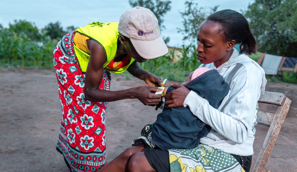
Then, ECG data acquisition was conducted using Lead II while the participants are in a conventional sitting ECG recording position. Appropriate techniques and strategies were applied to improve signal quality, electrode skin interface conductivity, and reduce artefacts. Standard recording methods were used to setup the device and subjects for recording. Near the electrode placement areas, watches and jewellery were removed. Alcohol was used to clean the electrode attachment skin sites. The subjects were seated upright and relaxed prior to recording. The typical lead II configuration was used to place the leads. The negative electrode was placed on the side of the palm of the right forearm above the wrist, the positive electrode on the interior left leg just above the ankle, and the reference electrode on the interior right leg just above the ankle. Similar protocols were used to record ECG data for all sessions. A total of 100 ECG signals with 1-min duration were collected from 50 participants (25 control and 25 experimental). The ECG signal acquisition procedure for the khat chewing portion of the study is displayed in Fig. 2.

ECG data acquisition procedure to investigate the effect of khat chewing on heart activity.
The effect of rehabilitation therapy from khat addiction on heart activity
For the rehabilitation therapy from khat addiction part of the study, data was obtained from khat addicts admitted to rehabilitation centres on the first day of admission and at the eighth day of admission for each subject. Most of the khat-addicted subject get recovered after a one-week stay in the rehabilitation program. In the rehabilitation centre, participants used to take medicines such as benzodiazepine, antidepressant or clonidine and therapies like watching TV, playing dart, playing table tennis, weight lifting, rope jumping and other sport activities as recommended by the physician. A case study approach was employed for this part of the study. The sample size was limited to 5 because most of the subjects with other co-addictions including alcohol, cigarettes, marijuana, opium, or a combination of one or more of these were excluded from the study. 10 ECG signals were recorded from these 5 subjects (1 female and 4 men), 5 before rehabilitation therapy and the remaining 5 after rehabilitation therapy.
Ethical approval
The study was approved by institutional review board of Jimma institute of health, Jimma university, with permission number JHRPGN/75/21, and institutional review board of Saint Paul’s hospital millennium medical college, with approval number PM23/385. In addition, an informed written consent was obtained from all study participants prior to data collection. All methods were carried out in accordance with the ethical standards as laid down in the 1964 Declaration of Helsinki and its later amendments or comparable ethical standards.
Study setting
For the effect of khat on heart activity part of the study, data collection for healthy control and chewer subjects was conducted in Jimma University Medical centre according to its intuitional protocol. For the effect of rehabilitation therapy from khat addiction on heart activity part of the study, data was collected in the psychiatric department of Saint Paul’s Hospital Millennium Medical College and Addis Hiwot rehabilitation centre according to their respective protocols.
ECG signal pre-processing
Following signal accusation, all the recorded ECG signals annotated and denoised. The recorded signals were given a unique name to identify the subject category, recording session, and counting number. Low-frequency noises caused by respiratory muscle movement, temperature change, and electrode motion artefact was removed. The Savitzky Golay filter was used to smooth the signal and remove low frequency disturbances20.
ECG signals are susceptible to disturbances such as powerline interference, EMG noise, and electromagnetic interference, in addition to baseline wandering abnormalities. As a result, any unwanted noises should be eliminated before extracting the relevant features. For our purpose, discrete wavelet transform which provides great performance for denoising ECG signals from these noises21,22,23 were used for signal denoising. Because the signals sampling frequency was 1000 Hz, the wavelet decomposition had frequency range patterns of 250–500 Hz, 125–251 Hz, 62.4–125 Hz, 31.2–62.6 Hz, 15.6–31.3 Hz, 7.79–15.7 Hz, 3.9–7.83 Hz, 1.95–3.92 Hz, 0.975–1.96 Hz, 0.487–0.979 Hz, 0.244–0.489 Hz, 0.0–0.243 Hz for detail (D) coefficients D1, D2, D3, D4, D5, D6, D7, D8, D9, D10, D11 and approximate (A) coefficient, respectively. Decomposition level 9 was used for eliminating frequency bands below 0.979 Hz that is assumed to be baseline wandering noise. The wavelet denoised signal was decomposed into wavelet coefficients and the energy of each coefficient was computed using a wavelet multiresolution analysis (MRA) technique to remove the residual noise. The low-frequency coefficients with a frequency range of less than 1 Hz which is out of the ECG signal range were excluded during wavelet reconstruction. Similarly, the frequency coefficients in ECG range having insignificant energy contribution were excluded during reconstruction. As a result, for reconstructing the denoised signal the contributor coefficients were selected from level 5 to level 9.
Feature extraction
The temporal peak detection and interval calculation techniques were used to calculate the time domain ECG characteristics, following the Pan Tompkins QRS detection24 approach. Pan Tompkins algorithm is a time-domain QRS detection algorithm that consists of a series of lowpass filter, high pass filter, derivative filter, squaring, thresholding, and moving windowing procedures. The heart rate is calculated from the detected R peaks of the QRS complex. Figure 3 shows the feature extraction model employed in this study.

Feature Extraction model.
MATLAB peak detector functions “max” and “min” were used for detecting the location and amplitude of maximum and minimum peaks with in the calculated temporal moving windows. The maximum and the minimum amplitude points in each moving window were detected as R peaks and S peaks respectively. For Q wave the temporal window was between the left margin of the moving window and the R peak location, for P wave between the left margin of the moving window and the Q peak location and for T wave between S peak and right margin location of the moving window. The intervals and segments were calculated from the onset and offset points of each waves. The HRV was calculated using root mean square of successive differences (RMSSD) between each R-peak. Finally, all the important calculated features were exported to an excel format from MATLAB workspace for further analysis.
Data analysis
The extracted features were averaged for better data manipulation. The changes between the averaged before and after chewing session for both chewers and controls were determined. Similarly, the changes between the averaged values before and after rehabilitation therapy were determined and the percentiles were computed from these values. In addition, the results have been statistically analysed using a pairwise t-test to show statistical differences among different groups (pre-chewing vs post-chewing of the experimental group, pre- vs post for the control group, pre-chewing of the experimental vs the control and post-chewing session of the experimental vs the control group).
Informed consent
An informed written consent form was obtained from all study participants.